Structures
Solved structures
Spatial structures of proteins and peptides solved in the laboratory by NMR (and by X-ray crystallography) and deposited in the Protein Data Bank. Images link to the RCSB PDB entries.
Receptors and membrane proteins
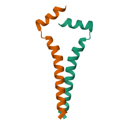
Transmembrane domain of the amyloid precursor protein (APP)
PDB 2LOH →
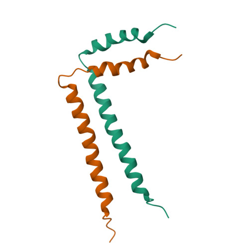
Transmembrane domain of the HER2 / ErbB2 receptor
PDB 2N2A →
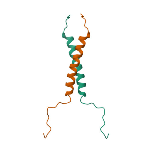
Transmembrane domain of the EGFR receptor (dimerisation)
PDB 5LV6 →
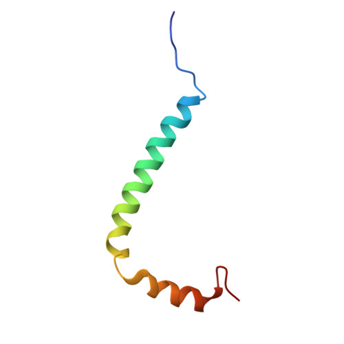
TM and juxtamembrane domains of EGFR
PDB 2N5S →
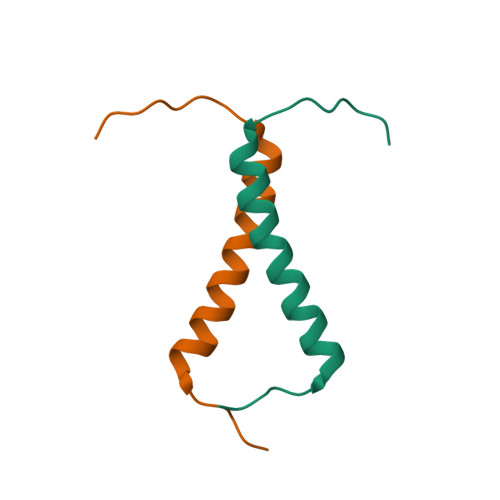
Transmembrane domain of the TrkA receptor
PDB 2N90 →
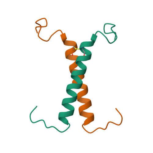
Transmembrane domain of the p75 receptor
PDB 2MIC →
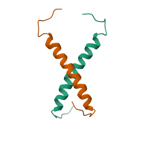
Transmembrane domain of the p75 receptor (C257A mutant)
PDB 2MJO →
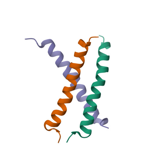
TM domain of toll-like receptor TLR3 (trimer)
PDB 2MKA →
Toxins and antimicrobial peptides
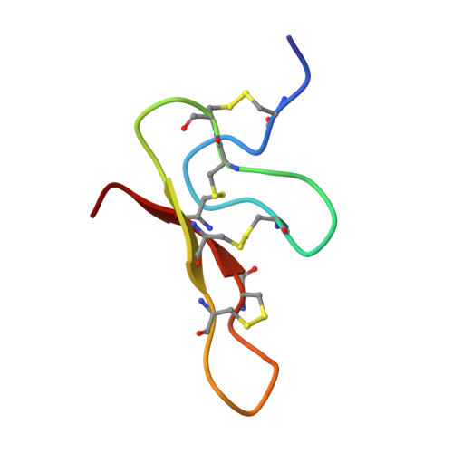
Purotoxin-1 — a neurotoxin from spider venom
PDB 2KGU →

Purotoxin-2 (in DPC micelle)
PDB 2MZG →
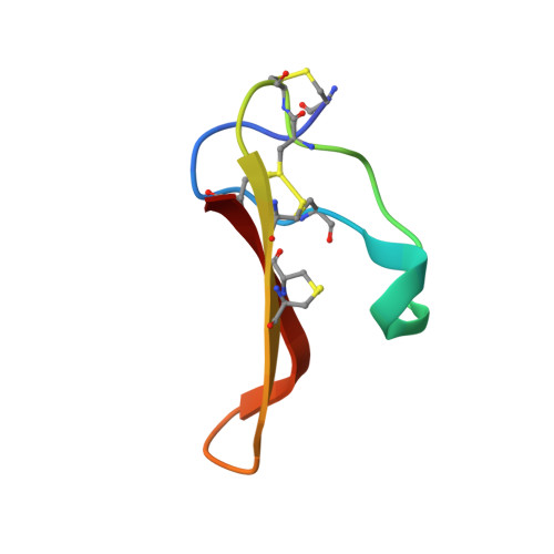
ω-Tbo-IT1 — insect Ca-channel blocker (spider venom)
PDB 2MYH →
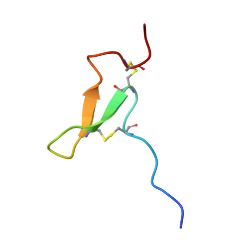
π-AnmTX Ugr 9a-1 toxin from sea anemone
PDB 2LZO →
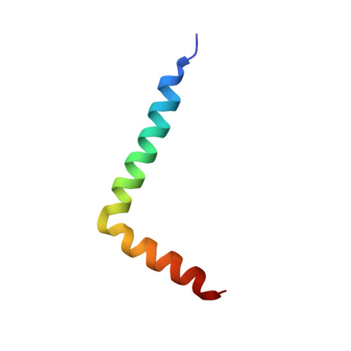
Antimicrobial peptide OtTx1a (in DPC micelle)
PDB 2N85 →
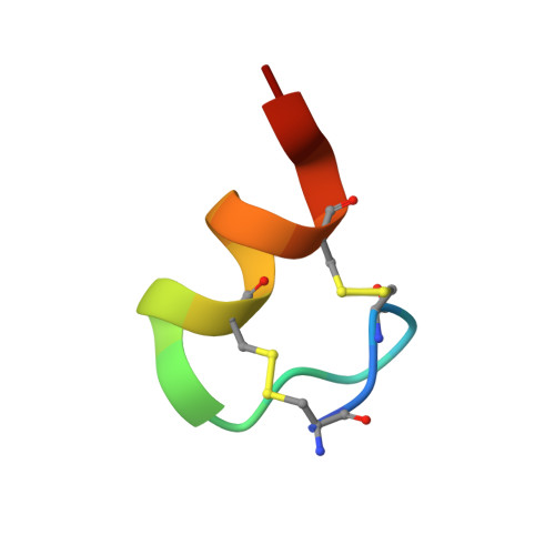
Apamin — a honeybee venom peptide
PDB 7OXF →

Antimicrobial peptide aurelin (from jellyfish)
PDB 2LG4 →
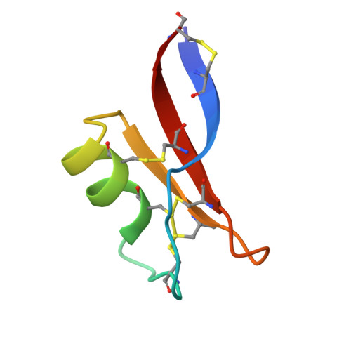
Plant defensin Lc-def
PDB 2LJ7 →
Other proteins

Exiguobacterium sibiricum rhodopsin
PDB 4HYJ →

FAST fluorescent reporter (dimer)
PDB 7AV6 →

Extracellular region of PsrSp (S. pneumoniae)
PDB 9A9G →
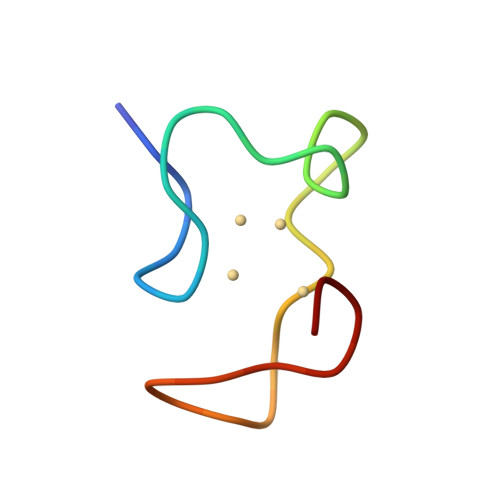
Rabbit liver metallothionein-2A
PDB 1MRB →
Structures deposited by members of the laboratory are shown. The full, up-to-date list is available in the Protein Data Bank and on the laboratory page at IBCh RAS.